Wound Care
There are many different types of wounds.
Those that require treatment have for whatever reason become problematic due to a break down in the healing process. Wounds such as leg ulcers, venous or arterial, diabetic foot ulcers, pressure sores, lymphatic skin conditions or injury sites that have become infected.
A vast majority can be accompanied, or a result of oedema. Oedema brings with it, bacteria, viruses and waste products that compounds the resistance to healing as well.
As mentioned on the Scar Tissue Therapy page, when an injury or wound occurs, the body goes through four stages of healing.
- Haemostasis – clot and stop the bleeding
- Inflammation / Immune response – clean up and remove waste & debris
- Proliferation – reconnect the damage fibers
- Remodeling – rebuild the structure
An interruption to this process is what causes a wound to become hard to heal or non-healing. 75-80% of the time, this occurs in stage 2, where the body is struggling with inflammation.
This is where Sal can help. First, she’ll take a medical history, account for any social, psychological and physical factors that may contribute to one’s capabilities, needs and wants, and of course assess the wound.
Wound healing is a complex and very fragile process. Failure to progress through the stages will likely lead to chronic and painful outcomes.
Evidence based wound care can speed up the stages of wound healing by keeping wounds moist, clean and protected from reinjury and infection.
Although the stages of wound healing are linear, wounds can move backward or forward depending on the patient, therapeutic, environmental and resource availability.
Risk of Infection
As mentioned on the Surgery Care page, open wounds, incisions or breaks in the skin is at risk of infection. It’s very important to keep the area clean, moist and protected from any friction or outside elements at least until the wound closes over.
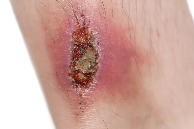
Signs of infection
- Heat
- Redness, watch for this expanding
- Mucus / slough
- Malodour
- Excessive fluid leakage past the 7-10 days post surgery
Please see your surgeon immediately or get to emergency, if any of the above are present.
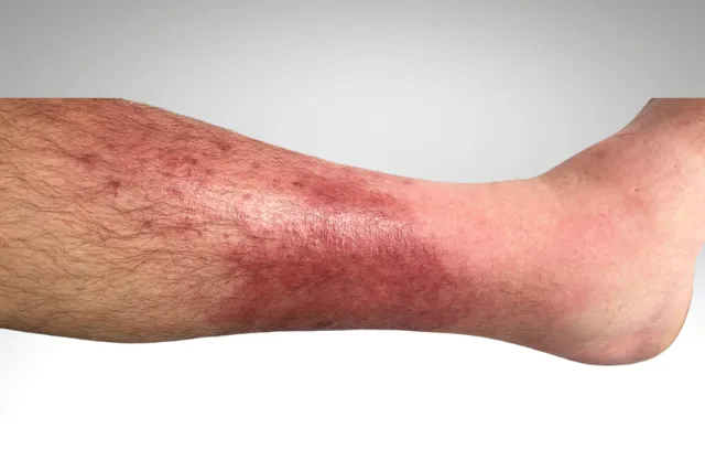
Risk of Cellulitis
Cellulitis is bacterial infection that affects the skin and tissue beneath it. It can develop anywhere on the body, but it’s most common on the lower legs and feet. Any sign of cellulitis, you must get yourself to emergency immediately, it can progress and become urgent very quickly.
Symptoms
- Redness, warmth, and swelling of the skin
- Tenderness or pain in the affected area
- Weeping or leaking of pus or clear fluid
- Fever and chills
- Blisters, spots, or dimpling of the skin
- Swollen glands in the armpit or groin.
Causes
- A break in the skin, such as a cut, wound, scrape, insect bite, or animal bite
- Skin conditions like eczema or fungal nail infections
- Ulcers from diabetes or other diseases
- Certain medications that suppress the immune system
- A recent surgery wound
Treatment
- Antibiotics are usually prescribed to treat cellulitis.
- Early treatment can prevent the infection from spreading.
- More serious cases may require hospitalisation.
Leg Ulcers
Lower limb wounds are the most concern due to high prevalence amongst those with impaired quality of life. Especially our seniors, diabetic sufferers, and those with chronic conditions.
There are many circumstances that may lead to lower limb wounds:
- Venous insufficiency (most common)
- Atherosclerotic arterial ischemia – Arterial flow issues
- Neuropathy – loss of sensation (diabetes, leprosy, chemotherapeutic, antibiotic induced)
- Lymphoedema
- Autoimmune conditions (rheumatoid arthritis, lupus, scleroderma)
- Infection (bacterial, fungal, parasitic, viral)
- Non-atherosclerotic ischemia (Martorell, vasculitis, sickle-cell disease)
- Inflammation (Pyoderma gangrenosum, Necrobiosis lipodica)
- Haematologic conditions (sickle-cell anaemia, anti-phospholipid syndrome)
- Malignancy (primary skin lesion, Marjolin)
- Medications (reactions, ulcer causing medications such as Hydroxyurea and Doxorubicin)
- Radiation fibrosis syndrome
- Trauma and bites
Most Common Leg Ulcers
Venous Ulcer – Those with Venous Insufficiency are at a high risk for ulcers, typically occurring on the lower leg, and in particular, around the medial ankle. The ulcer is often hard-to-heal, due to inadequate blood flow, high venous pressure and persistent inflammation – related to ‘iron-loading’ of the skin and the predisposition to biofilm (germ) invasion.
Arterial Ulcer – Arterial insufficiency impairs wound healing as tissues are deprived of oxygen and nutrients due to poor arterial blood flow and the delivery of immune cells is often compromised. Arterial ulcers are the second most common type of lower limb wounds, corresponding to 10–25% of cases.
Risk factors for atherosclerosis include:
- Family history of myocardial infarct/stroke
- >50 years of age
- Overweight
- Sedentary
- Smoking
- Diabetes
- High blood pressure
- Abnormal LDL/HDL profile
Diabetic Foot Ulcer – Diabetic foot ulcers are one of the most common complications of individuals who have uncontrolled diabetes. The ulcers result from poor glycaemic control, underlying neuropathy, peripheral vascular disease, or poor foot care.
The development of a diabetic foot ulcer generally occurs in three stages:
- A callus develops often as a result from neuropathy.
- Ongoing trauma occurs due to motor neuropathy causing physical deformity of the foot, sensory neuropathy causing loss of protective sensation, and drying of the skin due to autonomic neuropathy.
- Repeated trauma of the callus leads to subcutaneous haemorrhage and erosion, resulting in an ulcer.
Diabetes (type 2 in particular) is associated with high blood pressure, high blood sugar, abnormal blood lipids, insulin resistance, high visceral adipose accumulation, elevated inflammatory markers, endothelial (cells) dysfunction and a prothrombic (calcification & clots) state, making your wound extremely hard to heal or even non-healable without making some big changes.
Lymphoedema Wounds & Skin Changes
Chronic inflammation that accompanies lymphoedema can cause changes in the thickness, texture and shape of the skin, which leads to an overgrowth of the surface layer, wounds.
Such changes are:
Fibrosclerosis – inflammation and fibrous lesions
Hyperkeratosis – thickening of the skin, due to excess keratin production
Pachydermia – thick, heavy, hard (elephantitis) skin
Lymphangiectasia – small, raised skin lesions that contain clear lymphatic fluid and appear as tiny blisters over lymphatic malformations. They can look like blood blisters and may turn dark purple or black if blood leaks into them
Papillomatosis – Small cysts, dry, wart-like spots on the ankles, feet, and toes. The skin may also thicken and develop folds and bulges
Lymphorrhea – Wet legs, lymph fluid leaking through the skin excessively, a big risk of developing a leg ulcer due to lymphorrea and maceration of the skin
Complications
Cellulitis – is a bacterial infection within the skin and underlying tissues. This is quite a dangerous infection that requires immediate medical attention and antibiotics.
Myocosis – contagious fungal infection
Malignant, secondary Tumors
Safe to say, all of the complications listed above, require medical attention first before any lymphatic treatment can go ahead.
Common Factors Inhibiting Wound Healing
Patient Physical & Social Factors – Age, Stress, Nutrition, Hydration, Mobility, Social Anxiety
Infection – halts the healing process in inflammation, then it’s a constant battle between good and bad bacteria. The winner takes all. Keeping your wound clean is extremely important.
Medications – Antibiotics, steroids, non-steroidal anti-inflammatory drugs, chemotherapy, hypertensives medications all have side effects that can hinder the wound healing process, dependent on dosage and duration.
Radiation – destroys everything in it’s path, treatment to revitalise the impacted tissue will help with wound healing.
Other pathologies – Ischemia (discussed further in the next section), Autoimmune diseases and Oedema
Arterial Blood Circulation Issues
To clean up and fight against the microbes and biofilms that take over and infect our wounds, we need the assistance of the lymphatic system to take the waste, inflammation and disease away from within.
However, if you have an arterial flow issue, treating the lymphatic system will also likely impact the already struggling arterial flow. You will possibly need to seek addition investigations before treatment can go ahead.
Your extremities, limbs, feet, toes, hands and fingers will display signs of this issue.
- cold feet and fingers
- pale / bluish colour
- pain at rest (achy, burning, cramping)
- numbness and weakness
- toenails and fingernail changes
- skin changes
Tests in clinic to confirm or persuade the need for specialist help.
- Buerger’s Test
- Pedal Pulses
- Capillary refill
- Temperature changes (cool in the extremities)
- Oxygenation
- Hair loss
- Shiny skin
- Dry skin
Should the above be present / positive and cause for concern, you will need to go back to your GP to be referred to a vascular specialist. Lack of arterial flow will most certainly impede the healing process of your wound/s.
Treatment for Wounds
After evaluating your history, social, psychological and physical limitations, we need to assess the body presentation, wound site, peri-wound and surrounding tissue.
- Body – look for signs of other conditions
- Measurements – to gauge progress
- Colour
- Exudate – fluid leakage
- Odour
- Position
- Pain scale / personal feeling scale
Once assessment is complete, we can now treat.
In order for any wound to begin to heal it needs to have clean (free from the bug), moist environment.
- If too moist or leaking in excess, it needs absorbents,
- If too dry we need to add moisture, topical creams gels (type will depend of evaluation on healing or non-healing
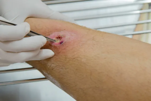
- Scabs, biofilm or mucus, necrotic tissue needs to be removed. To do this we may need to use a solvent called ‘surfactant’ this will help to separate the infected tissue from the base underneath.
- In some cases, after softening and separating we may need to scrub the area, to try and remove any attached biofilm from the new tissue. This can be painful, so care must be taken to adequately dissolve, gently scrub, or use a particular dressing to help trap and kill the bacteria. Topical anaesthetic creams can be used to reduce the discomfort.
- Dressing to be applied. There are way to many types of dressings to mention, however they each have an individual purpose.
As the wound progresses through the stages, the dressing type can also change. Always best to seek advice from a wound care therapist / nurse.
As for treatment frequency, no known treatment times can be quoted. It depends on home care and compliance. Getting on top of it as quickly as possible will ensure the best and fastest possible outcomes.
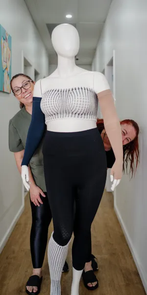
Compression Garments
The gold standard of care for wounds is compression.
Doing the very thing I have talked in length about throughout this entire website. Lymph drainage to flush the bacteria and germ away.
You will likely be bandaged in the interim, followed by a compression garment if necessary to maintain a lowered fluid level. See compression garments page for details under venous insufficiency.